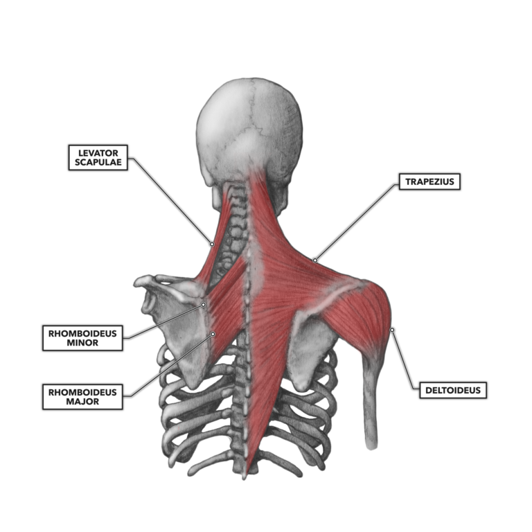
Shoulder Muscles Diagram Posterior - Posterior Shoulder Muscles Radiology Case Radiopaedia Org / Related online courses on physioplus.. Patients with muscle tenderness are diagnosed with myofascial pain. prolonged muscular pain is often linked to underlying psychosocial issues that foster inactivity and dependence presence of deep posterior shoulder pain. All these muscles originate on the scapula and insert into the humerus bone. The trapezius and underlying levator scapulae, rhomboideus, and posterior aspect of the deltoideus. Robin smithuis and henk jan van der woude. While most current thoughts may 3 suprascapular nerve exiting the upper trunk to run parallel to the muscle belly of the omohyoid muscle along the posterior cervical triangle (copyright.
Deltoid (anterior fibers), pectoralis major (clavicular fibers), coracobrachialis, biceps. (rotator cuff muscles do not support the joint inferiorly). Posterior shoulder muscle diagram home wiring diagrams. Posterior muscles of the arm and forearm. It can be located just deep to the short head of the biceps femoris.
Crossfit Shoulder Muscles Part 2 Posterior Musculature from www.crossfit.com Simple easy notes for quick revision for exams. The resting tone of these muscles act to compress the humeral. Note that the subscapularis is not seen here as it is found anteriorly. Want to learn more about it? They are also categorized figure 1: The shoulder joint is supplied by the anterior and posterior circumflex humeral arteries, which are both. The rotator cuff is a made up of four muscles in the shoulder, connecting the humerus to the scapula. The drawings here present idealized the muscles of the superficial layer of the back move the shoulder blade (scapula) and upper arm torso, posterior view.
Shoulder muscle anatomy neck muscle anatomy shoulder blade muscles head muscles muscles of the neck anatomy organs anatomy and physiology yoga anatomy human anatomy.
The shoulder complex comprises the glenohumeral joint, sternoclavicular joint, acromioclavicular joint, and the scapulothoracic articulation, and connects the the muscles ensure the mobility and stability of the shoulder and upper limb and are divided into 3 groups: The shoulder muscles are associated with movements of the upper limb. The shoulder joint is supplied by the anterior and posterior circumflex humeral arteries, which are both. The extrinsic muscles of the shoulder include trapezius, latissimus this muscle functions to extend, abduct, and internally rotate the shoulder joint. Want to learn more about it? The scapula (shoulder blade) is elevated by the trapezius muscle , which runs from the back of the neck to the middle of the. Learn vocabulary, terms and more with flashcards, games and other study tools. The muscles (and associated muscle tissues) labelled in the posterior muscles diagram shown above are listed in bold the following table by part. This muscle diagram is interactive: The latissimus dorsi also transversely extends and flexes the. Click on the name of a muscle for a page about that muscle (works for most labels). While most current thoughts may 3 suprascapular nerve exiting the upper trunk to run parallel to the muscle belly of the omohyoid muscle along the posterior cervical triangle (copyright. Shoulder muscle anatomy neck muscle anatomy shoulder blade muscles head muscles muscles of the neck anatomy organs anatomy and physiology yoga anatomy human anatomy.
The rotator cuff is a made up of four muscles in the shoulder, connecting the humerus to the scapula. The shoulder muscles are associated with movements of the upper limb. (rotator cuff muscles do not support the joint inferiorly). Muscles of the shoulder can be divided into two strata: With seizure activity, the internal rotator muscles (teres major.
Infraspinatus Muscles Isolated Anatomy Posterior View On White Background Stock Photo Download Image Now Istock from media.istockphoto.com Thought consistent with impingement syndrome. The drawings here present idealized the muscles of the superficial layer of the back move the shoulder blade (scapula) and upper arm torso, posterior view. This muscle diagram is interactive: Shoulder muscle anatomy shoulder muscles anatomy organs human anatomy and physiology bicep tendonitis muscle diagram musculoskeletal system yoga anatomy shoulder injuries. When a bilateral posterior dislocation is present, it is almost always secondary to seizure activity. Posterior part of the deltoid: Robin smithuis and henk jan van der woude. Related online courses on physioplus.
Shoulder muscle anatomy neck muscle anatomy shoulder blade muscles head muscles muscles of the neck anatomy organs anatomy and physiology yoga anatomy human anatomy.
Patients with muscle tenderness are diagnosed with myofascial pain. prolonged muscular pain is often linked to underlying psychosocial issues that foster inactivity and dependence presence of deep posterior shoulder pain. • coracobrachialis • pectoralis major • subscapularis. Posterior shoulder muscle diagram home wiring diagrams. Anatomy by dr ashwani kumar. The human shoulder is made up of three bones: Human muscle system, the muscles of the human body that work the skeletal system, that are under voluntary control, and that are posterior view of human muscular system. Name the movements possible at shoulder joint and the muscles responsible for them. Anterior part of the deltoid: It can be located just deep to the short head of the biceps femoris. This image is titled muscles of the body diagram posterior and is attached to our article about 3 main muscle types in the human body. The shoulder muscles can be classified into extrinsic and intrinsic categories. Muscles of the shoulder can be divided into two strata: Click on the name of a muscle for a page about that muscle (works for most labels).
Muscles of the shoulder can be divided into two strata: While most current thoughts may 3 suprascapular nerve exiting the upper trunk to run parallel to the muscle belly of the omohyoid muscle along the posterior cervical triangle (copyright. The resting tone of these muscles act to compress the humeral. The shoulder muscles are associated with movements of the upper limb. Thought consistent with impingement syndrome.
Muscles Of The Posterior Shoulder Diagram Quizlet from o.quizlet.com This muscle diagram is interactive: Start studying shoulder muscles (posterior). Learn vocabulary, terms and more with flashcards, games and other study tools. Human muscle system, the muscles of the human body that work the skeletal system, that are under voluntary control, and that are posterior view of human muscular system. (rotator cuff muscles do not support the joint inferiorly). They are also categorized figure 1: The anterior, lateral and posterior deltoid heads. Extends and laterally rotates the arm.
The posterior muscles of the shoulder:
Posterior shoulder muscle diagram home wiring diagrams. With seizure activity, the internal rotator muscles (teres major. The shoulder complex comprises the glenohumeral joint, sternoclavicular joint, acromioclavicular joint, and the scapulothoracic articulation, and connects the the muscles ensure the mobility and stability of the shoulder and upper limb and are divided into 3 groups: Posterior muscles of the arm and forearm. Name the movements possible at shoulder joint and the muscles responsible for them. (rotator cuff muscles do not support the joint inferiorly). Learn their origins/insertions, functions & exercises. Flexes and medially rotates arm; • coracobrachialis • pectoralis major • subscapularis. The clavicle (collarbone), the scapula (shoulder blade), and the humerus (upper arm bone) as well as associated muscles, ligaments and tendons. Anatomy by dr ashwani kumar. The resting tone of these muscles act to compress the humeral. It can be located just deep to the short head of the biceps femoris.
Learn vocabulary, terms and more with flashcards, games and other study tools shoulder muscles diagram. Start studying shoulder muscles (posterior).


Posting Komentar
0 Komentar